
The Wound Care Pathway: Your 5 step guide to wound healing
PDF
6MB
25
pages
By following the steps in this pathway, you can provide an optimal healing environment for skin tears and reduce the risk of complications that could lead to delayed healing or worse.
Any advice included here needs to work in conjunction with your local protocols and your individual scope of practice.
This article is based on the book A pathway for treating a person with a: Skin tear.
A skin tear is a wound caused by shear, friction and/or blunt force resulting in separation of skin layers. A skin tear can include partial thickness or the full thickness of the skin.1
Skin tears are traditionally categorised as acute but without the appropriate treatment they can become chronic.
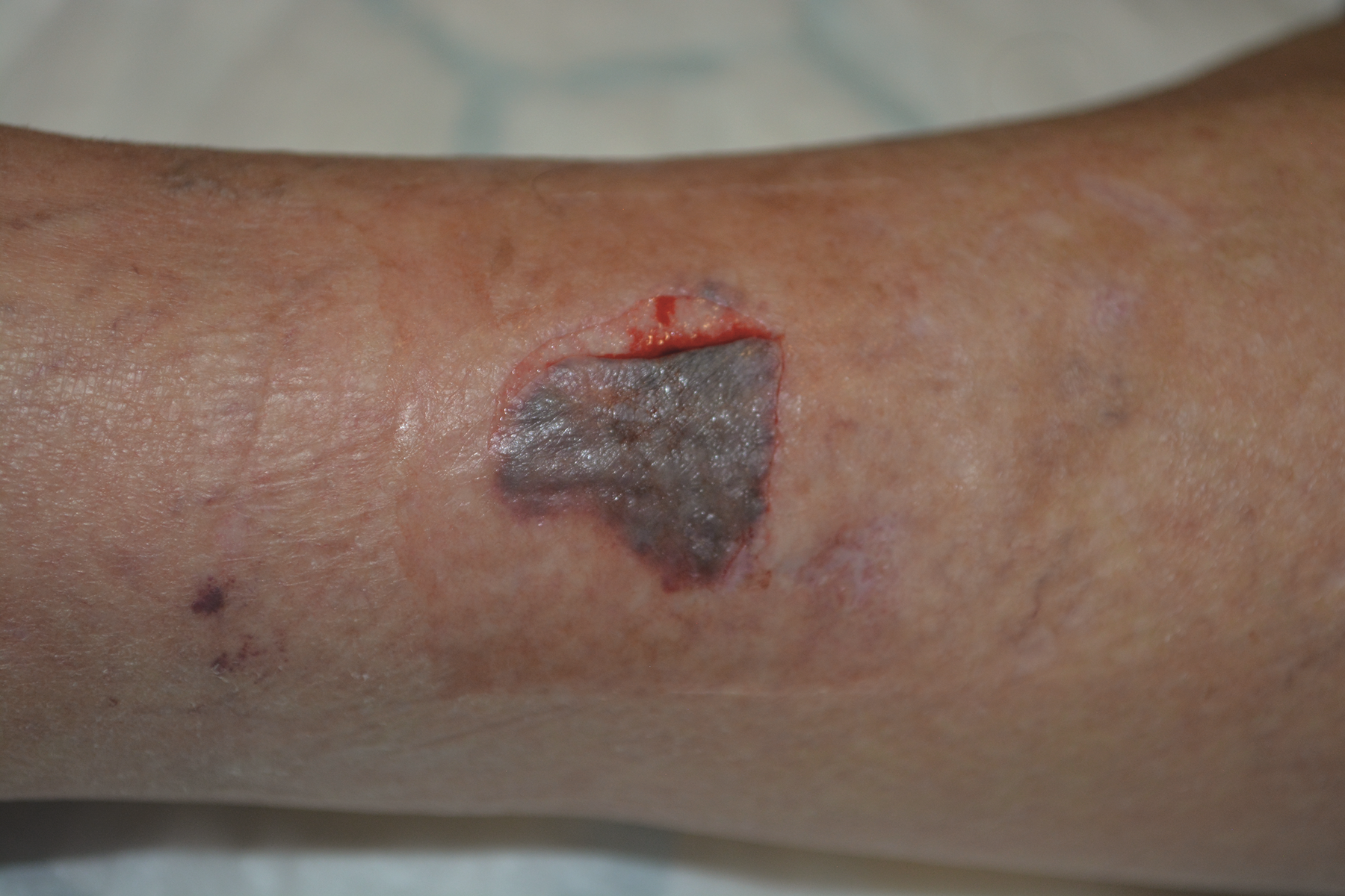
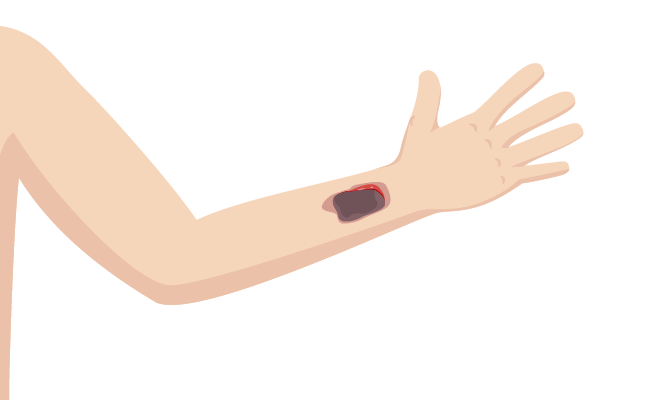
Type 1 skin tear
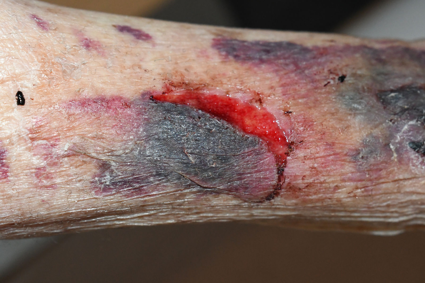
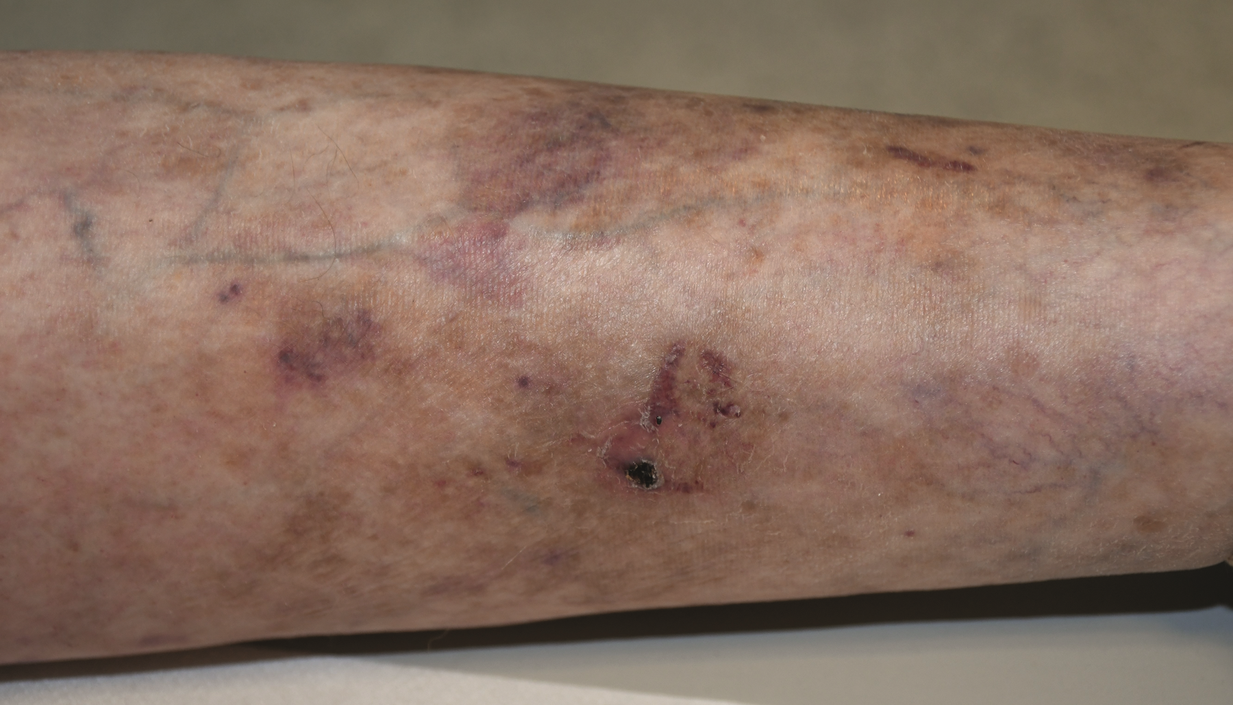
Type 2 skin tear on the lower leg
The guidance provided in this Wound Type Specific Pathway, is best understood in combination with the detailed guidance available to you in The Wound Care Pathway.
➔ Begin your holistic patient and wound assessment, to determine skin integrity. Several factors need to be taken into account:
➔ Explore the cause of the skin tear:
If it was caused by an external factor then screen for safety factors.
➔ Assess and administer first aid if necessary, and determine if Tetanus vaccination is required (recommended for patients who have not received Tetanus during the last 10 years).
➔ Assess blood loss and determine if the patient is on anti-coagulants.
➔ Screen for associated fractures, especially if the patient fell, etc.
In all wound types and skin conditions, it is important to be aware of how signs and symptoms may present in a range of skin tones.2
➔ In order to diagnose a skin tear, you first have to classify the wound based on ISTAP classification system: 3,4

Type 1: No skin loss where flap can be repositioned to cover the wound bed
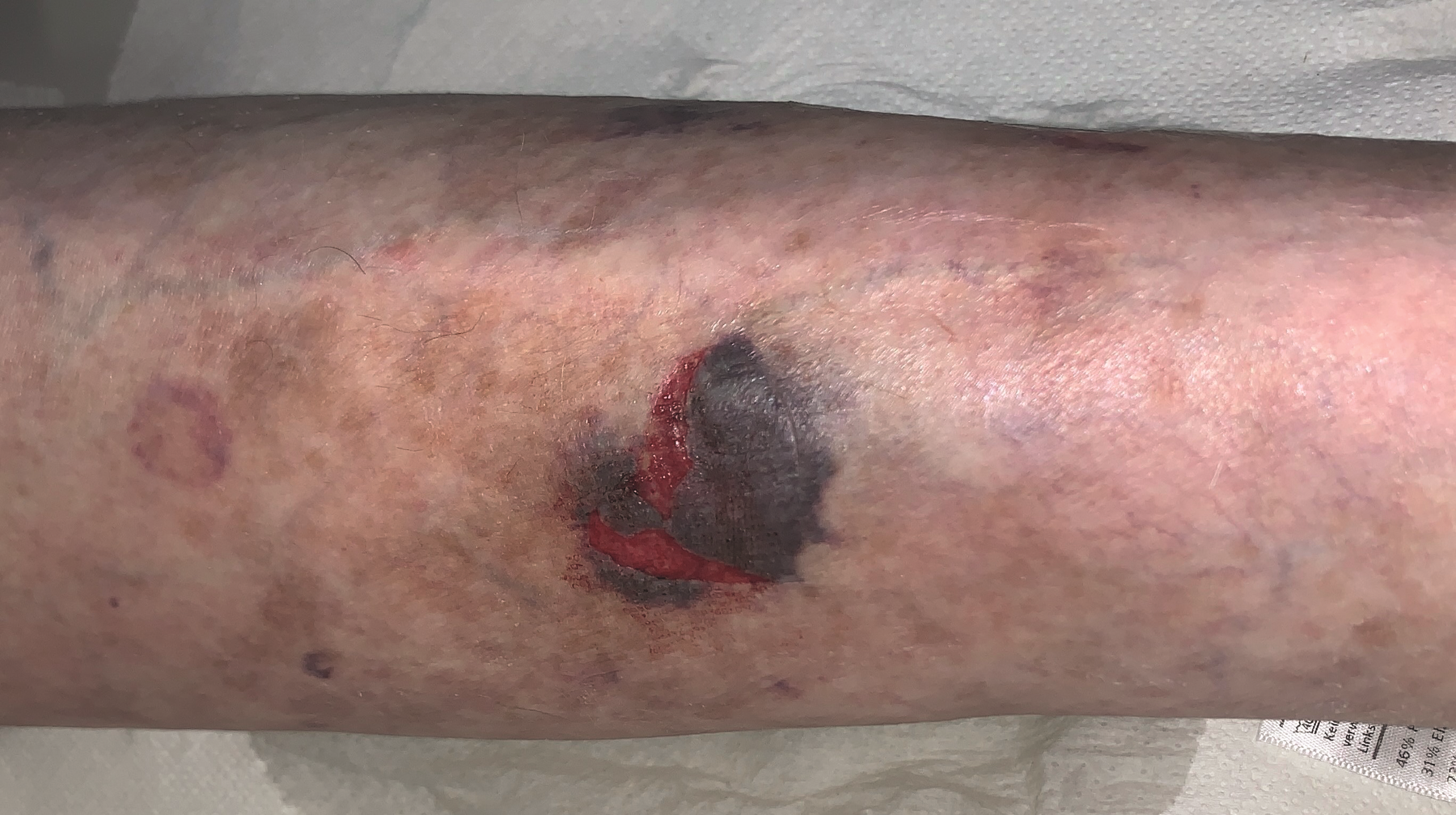
Type 2: Partial flap loss where flap can be repositioned to cover the wound bed
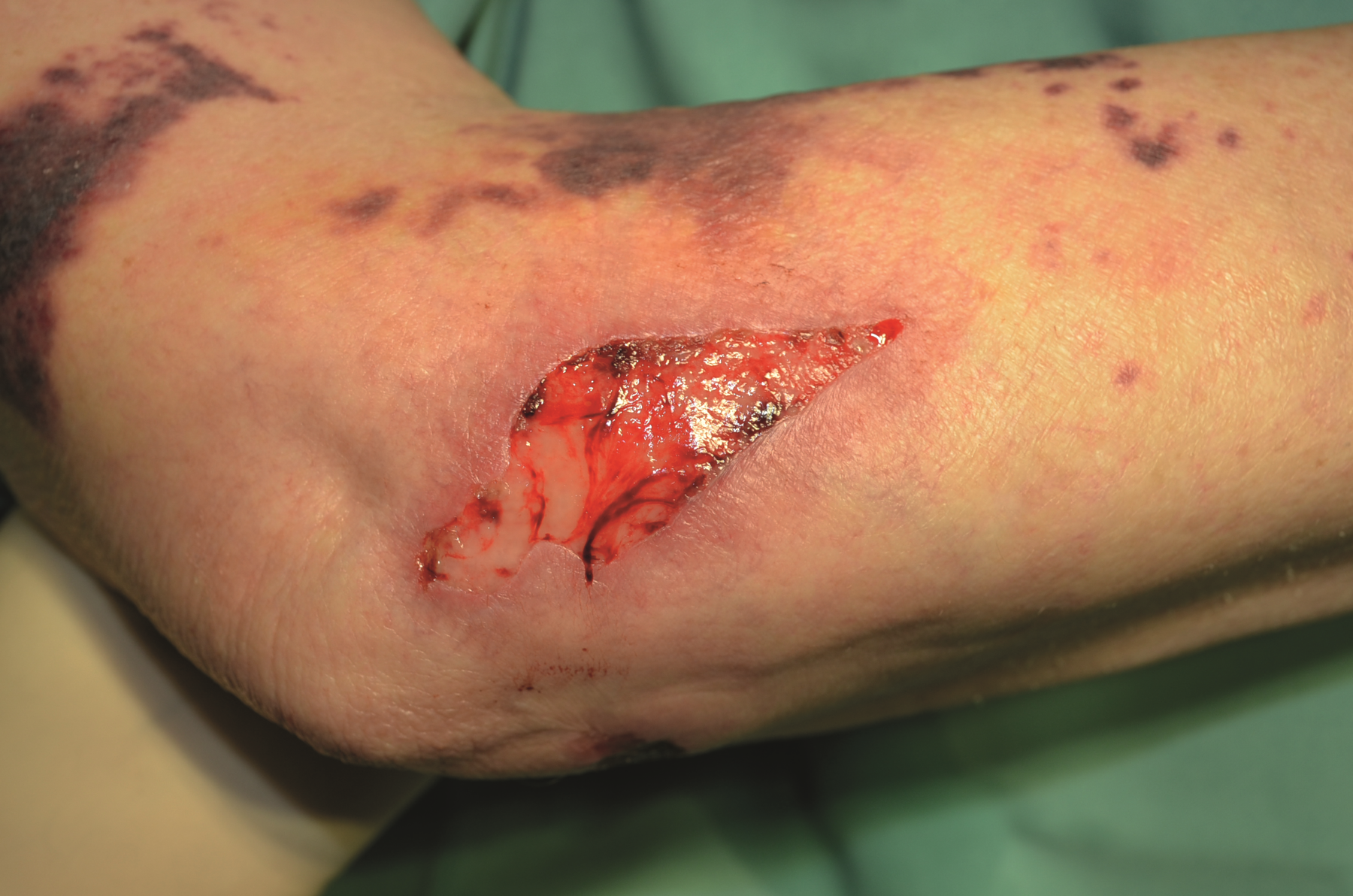
Type 3: Total flap loss exposing the entire wound bed
➔ Proceed by making an assessment about the duration of the skin tear:
➔ You also need to measure and document the wound size
➔ Next, assess the wound bed characteristics
➔ Finally, consider the location of wound and assess its implications:
For more information, view the ISTAP best practice recommendations.
➔ Develop your treatment plan taking the age of the wound into consideration – as well as several other factors.
➔ Consider pain control.
➔ Consider skin integrity, and the need for nutrition and hydration – moisturizing twice daily is recommended.5
➔ Develop prevention protocols to manage risks – environmental hazards and infection risk
Associated large haematomas / deep dissecting haematomas should be evacuated or referred. You can determine the depth and extent of a haematoma by palpating the area, assessing the range of motion, assessing distal circulation and identifying severity of pain.
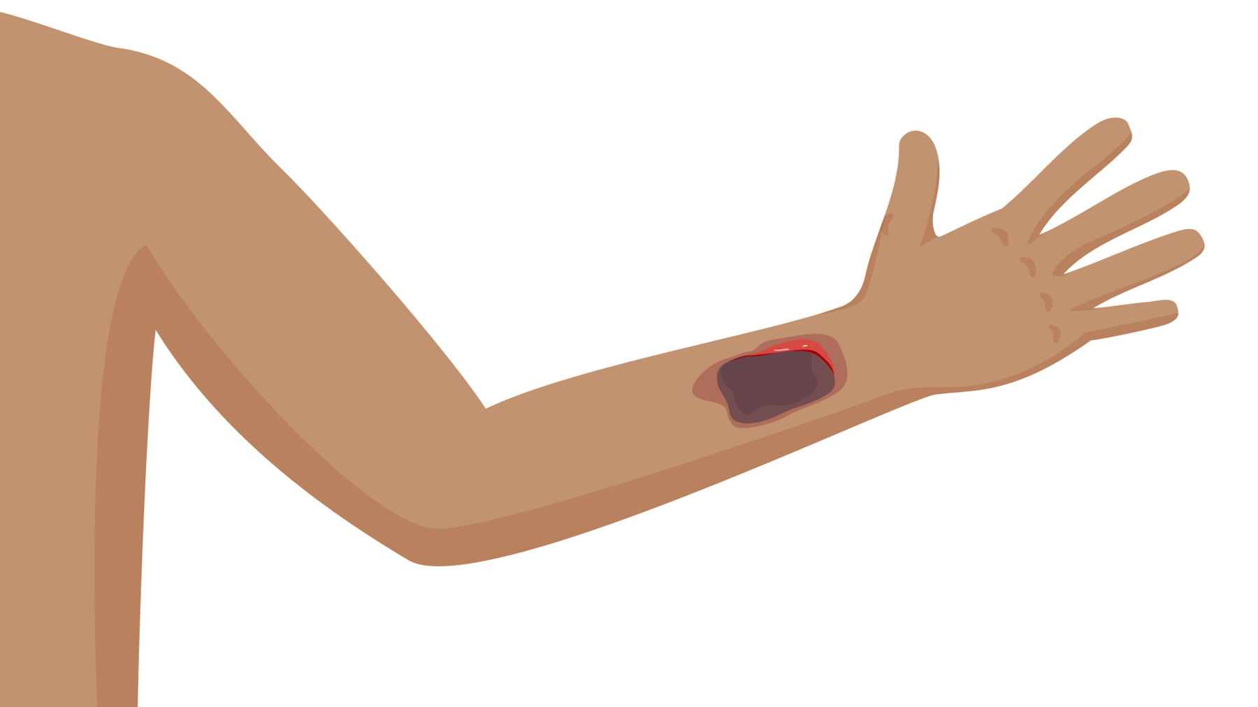
Large haematoma with suspected extension
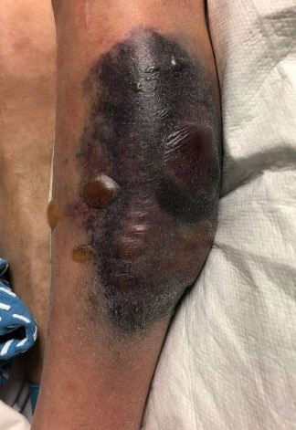
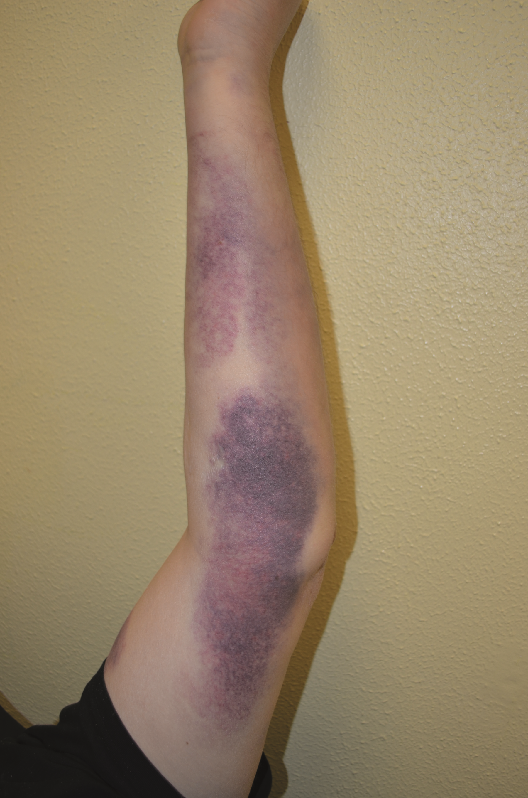
Large haematoma restricting blood flow, cousing local oedema and inflammation
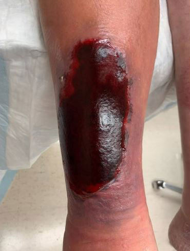
Large haematoma requiring evacuation
Discover our free online medical education programme supporting clinicians who are managing wounds and skin care complications.
In case you are dealing with a new skin tear (at the time of injury):
➔ Administer first aid.
➔ Stop bleeding with gentle pressure.
➔ Clean wound with non-irritating cleansers, potable water or saline.
➔ Re-approximate the skin flap by gently rolling the flap back into place using dampened sterile cotton tip applicators, gloved fingers or sterile tweezers/forceps.7
➔ Check flap after 24 hours for type 1 and 2.
Suturing of skin tears is not generally recommended.8 And the maximum length to width ratio of a skin flap on the extremities should be 1:1, flaps beyond this have a higher risk of failure.9
If you are using adhesive strips, then position the strips apart and allow them to fall off. Do not remove them!
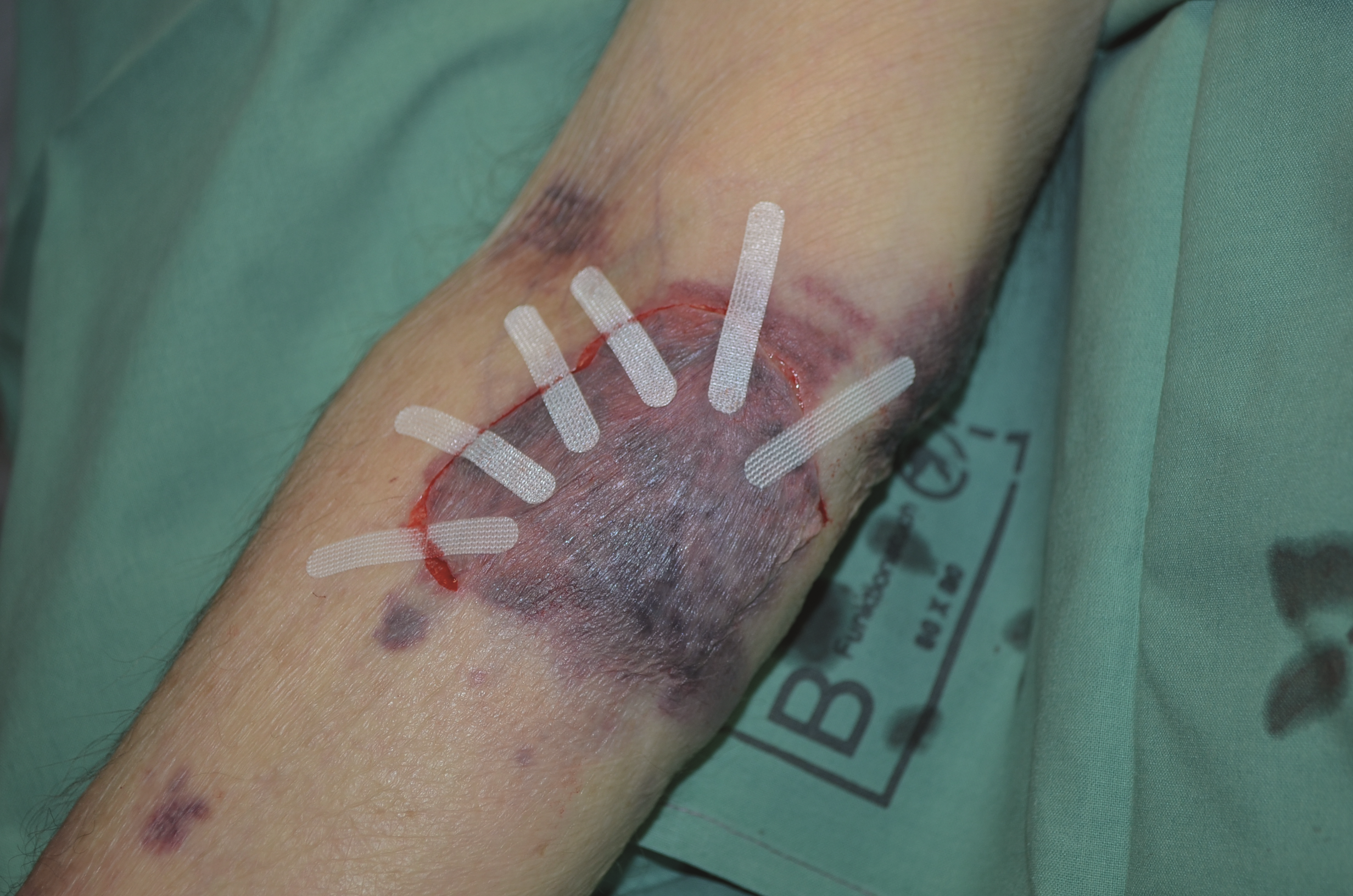
Positioning of adhesive strips
In case you are dealing with an older skin tear:
➔ Clean wound with saline.
➔ Debride non-viable tissue.
➔ Watch/observe for signs of infection.
➔ Use topical antimicrobials in case of local infection and systemic antibiotics in case of spreading infection.
➔ Implement prevention protocols
➔ Always use a dressing that is appropriate for the level of exudate, the size of the skin tear and the skin type.
➔ Select a dressing that will be atraumatic upon removal, and will not cause any further damage to the wound bed and/or any remaining skin flap or the periwound skin.4
Avoid iodine-based dressings (drying affect) and film/hydrocolloid dressings (strong adhesives).
➔ Make sure the dressing facilitates moisture balance and protects periwound skin. Generally, skin tears are not heavily exudating wounds; however, in some cases, depending on the location and co-morbidities such as peripheral oedema, skin tears may be heavily exudating. Choose a dressing accordingly.
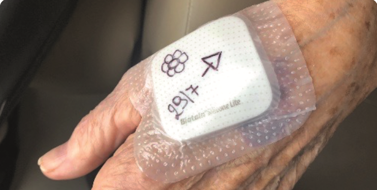
➔ Always draw an arrow on dressings to indicate correct direction of removal and write either the date for review or the date dressed.Minimize trauma by slowly removing dressing in the direction of the arrow, adhesive removers can also be used.
Compression therapy should be considered as an additional therapy if the wound is on an extremity. (Before applying compression on a lower leg, a full leg assessment including vascular assessment should be carried out.10 Light compression or support can be considered for an arm.)
➔ Conduct regular reassessments at time intervals that are appropriate for the severity of wound. A skin tear should not take more that 4 weeks to heal.
Healed skin tear
➔ Assess risk factors, including screening for co-morbidities which can increase the risk of chronicity (i.e. peripheral arterial disease, venous insufficiency, etc.)
➔ Discuss prevention strategies with patients based on risk factors.11
Extra care should be taken to avoid Medical Adhesive Related Skin Injuries (MARSI), as they can affect skin integrity, cause pain, increase risk of infection and potentially increase wound size and delay healing.(12) MARSI can be hard to identify when it occurs in patients, especially those with dark skin tones.(2)
– is an abnormal collection of blood outside of a blood vessel, causing swelling. A bruise is bleeding under the skin without swelling. The skin over a haematoma often feels spongy, rubbery and lumpy. Severity of haematoma depends on the size and depth. Refer patients if the haematoma is large, tense, painful, infected, over a joint or airway or is expanding.
- is a narrowing or blockage of the vessels that carry blood from the heart to the legs.
– is a condition in which the veins fail to return blood efficiently to the heart. Symptoms include swelling of the legs and pain in the extremities.
– stands for Medical Adhesive Related Skin Injury. It occurs when superficial layers of skin are removed by medical adhesive, resulting in skin trauma such as formation of vesicles, bulla, skin erosion, and skin tears, that persist longer than 30 minutes after removal of the adhesive.
For a glossary of general wound care terms consult The Wound Care Pathway: