
Get full access with a free account
Benefits of the Coloplast® Professional Educational platform
- Get full access to all educational content, events and resources
- Track your progress
- Share content with your collegues
- Share supporting material with your patient
Incorporating the aSSKINg Framework in Wound Assessment
When conducting an initial wound assessment, healthcare professionals should address the following elements:
-
Assess Risk
Begin by identifying factors that increase a patient’s risk of developing pressure ulcers. Use validated tools, like the Braden or Waterlow scales, alongside clinical judgment. Assess mobility, medical history, and external factors contributing to skin vulnerability. -
Skin Assessment and Care
Examine the patient’s skin, focusing on areas prone to pressure damage, such as bony prominences. Document any signs of erythema, moisture-associated damage, or early skin breakdown. Early detection during this step can prevent wound deterioration. -
Surface Selection
Ensure the patient is positioned on an appropriate support surface. Pressure-relieving mattresses, cushions, and overlays should be matched to the individual’s needs. Consider repositioning schedules to alleviate pressure on high-risk areas. -
Keep Moving
Encourage patients to reposition frequently to minimize prolonged pressure on specific areas. For immobile patients, establish a repositioning schedule tailored to their needs, such as using a 30-degree tilt technique. -
Incontinence and Moisture Management
Moisture from incontinence, sweat, or wound exudate can contribute to skin maceration and ulceration. Use barrier creams and specialized products to keep the skin clean and dry, protecting it from further damage. -
Nutrition and Hydration
Nutritional deficiencies and dehydration can impair skin integrity and delay healing. Assess dietary intake and, if needed, involve a dietitian to optimize nutrition and hydration plans. -
Giving Information
Educate patients, caregivers, and family members on wound prevention strategies, including repositioning techniques, equipment use, and the importance of adequate nutrition. Effective communication is essential to ensure compliance and successful wound management.
Risk Assessment Tools
Successful Pressure Ulcer prevention requires individual Pressure Ulcer risk profiling, including:3.
- Skin assessment
- Holistic assessment of the individual
The most frequently used risk assessment tools are:
- Purpose T: Best for simplifying risk assessment and linking directly to interventions; recommended by the National Wound Care Strategy Programme.
- Waterlow Scale: Offers a detailed, multifactorial approach but may require clinical judgment to avoid overestimations.
- Braden Scale: A globally accepted tool with a strong evidence base, excellent for proactive care planning.
Skin assessment frequency
NPUAP/EPUAP/PPIA guidelines recommend that skin assessments are conducted1:
- Within 6 hours of admission2 or on first visit/clinic appointment
- As part of every risk assessment
- Ongoing based on the clinical setting and the individual’s degrees of risk
- Prior to discharge from caseload
- Increase frequency in response to any deterioration in overall condition
The skin assessment should be a head-to-toe assessment with focus on skin overlying bone prominence including the sacrum, ischial tuberosities, greater trochanters and heels.
At risk areas
|
|
The difference between a Pressure Ulcer and MASD
| Pressure Ulcer | MASD (incl moisture lesions) | |
|---|---|---|
|
Cause |
Pressure and/or shear forces internal or external |
Moisture( MASD): incontinence (incontinence associated dermatitis), sweat( intertrigininous dermatitis/Intertrigo, peristomal(stoma), periwound(wound exudate and friction |
|
Location |
Usually bony prominence, occuring in a defined area |
Sacrum, perineum, stoma, wound edges, skin folds, any part of the body exposed to moisture |
|
Depth |
Superficial to full thickness |
Superficial to partial thickness |
|
Shape |
Defines edges, often circular or irregular shape |
Diffuse area (moisture uncontained). ‘Kissing’ ulcers may be present |
|
Necrosis |
Possible necrosis |
No necrosis |
|
Colour of wound bed |
Non blanching erythema, Slough, Necrosis |
Red, pink or white. Not equal |
The Role of the aSSKINg Bundle in Initial Wound Assessment
Effective wound care begins with a thorough initial holistic assessment, ensuring that the wound’s condition and contributing factors are fully understood. The aSSKINg bundle offers a systematic approach that healthcare professionals can incorporate into their initial wound assessment process. This structured framework ensures comprehensive evaluation and optimal care planning, reducing the risk of complications such as pressure ulcers.
Category 1 — Non-blanchable erythema (intact skin)
)
- What it is: Skin is unbroken with a red area that doesn’t turn white when pressed.
- In darkly pigmented skin: May appear darker in colour, check for temperature changes, oedema, changes in tissue consistency and pain
- Why it matters: First sign of pressure damage.
- Key point: Skin not open or broken.
Category 2 — Partial-thickness skin loss
)
- What it is: Partial thickness loss of the dermis — may look like a shallow open wound or clear fluid filled blister.
- Key point: if broken dermis exposed, but fat not visible, slough and necrosis not present.
Category 3 — Full-thickness skin loss
)
- What it is: Full thickness tissue loss, with adipose (fat) visible.
- Key point: No bone, muscle, tendon, or cartilage exposed.
Category 4 — Full-thickness tissue loss
)
- What it is: Full thickness skin and tissue loss, Deep wound with exposed bone, tendon, muscle, or other deep structures.
- Key point: Indicates most severe tissue damage, potential risk of osteomyelitis
Unstageable Pressure Ulcer
)
- What it is: An ulcer where the true depth can’t be determined because slough (yellow/tan) or necrosis (brown/black) obscures the wound bed.
- Key point: Once slough/necrosis is (safely) removed, wound usually reveals a Stage 3 or Stage 4 pressure ulcer and should then be categorised accordingly
Deep Tissue Injury (DTI)
)
- What it is: Intact skin, broken skin or blood-filled blister with persistent deep red, maroon, or purple discoloration, often from damage under the skin due to pressure/shear.
- Key point: Wound can evolve rapidly
Assessment and pressure ulcer location
Some locations have multiple areas where damage could occur e.g. buttocks and feet.
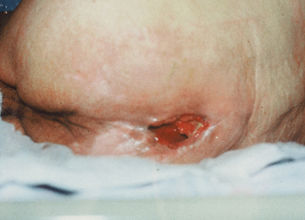
Pressure Ulcer on the lower back.
Copyright of Brenda King
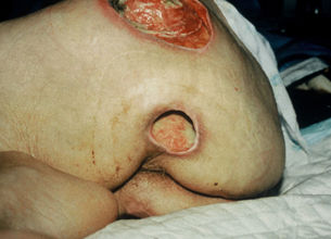
Pressure Ulcer on the left buttock and upper leg.
Copyright of Brenda King
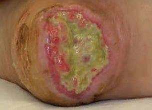
Pressure ulcer on a heel
Be aware
Be aware of skin changes at end of life (SCALE) as part of the holistic patient palliative care plan. One example are Kennedy ulcers : A dark sore that develops rapidly during the final stages of a person’s life. Kennedy ulcers grow as skin breaks down as part of the dying process. Not everyone experiences these ulcers in their final days and hours, but they’re not uncommon.4
How to effectively prepare Pressure Ulcers
- First, you should clean the wound and surrounding skin with low-toxicity solutions such as potable/sterile water or sterile saline
- If infection is suspected, the use of antiseptic solutions for cleaning the wound and periwound is recommended
- Remember: Irrigation of the wound should be avoided, if you can not see where the irrigation solution is going. Compresses can be used if irrigation is not possible
- Debride the wound, using Alprep Pad, to remove non-viable tissue unless there is stable eschar on a heel or an area of poor perfusion
- If you assess that you are dealing with an infection, proceed according to the IWII Infection Continuum & Management Guide
- Pay close attention to the wound’s moisture/exudate balance

It's not what we put on the wound that heals it; it's what we take off
Podiatry Operations Manager
Considerations when treating patients with Pressure Ulcers
First determine if you are dealing with a pressure injury or something else. Make your assessment of the skin or wound using the NPIAP/EPUAP/PPIA classification system.
Identify goals based on healability of the wound. Goals may include: decrease in wound size, alleviation of smell, decreased discharge and pain, etc.
Category 1 — Non-blanchable erythema (intact skin)
How to treat: Off load pressure, no dressing required, follow your local guidance
- What it is: Skin is unbroken with a red area that doesn’t turn white when pressed.
- In darkly pigmented skin: May appear darker in colour, check for temperature changes, oedema, changes in tissue consistency and pain
- Why it matters: First sign of pressure damage.
- Key point: Skin not open or broken.
Category 2 — Partial-thickness skin loss
How to treat: Intact fluid filled blister: off load pressure, no dressing required, follow your local guidance
- What it is: Partial thickness loss of the dermis — may look like a shallow open wound or clear fluid filled blister.
- Key point: if broken dermis exposed, but fat not visible, slough and necrosis not present.
Category 3 — Full-thickness skin loss
How to treat: Open wound: Wound prep, comfeel transparent / plus or Biatain Silicone
- What it is: Full thickness tissue loss, with adipose (fat) visible.
- Key point: No bone, muscle, tendon, or cartilage exposed.
Category 4 — Full-thickness tissue loss
How to treat: Up to 2cm in depth, wound preparation using Alprep Pad, Biatain Silicone. Over 2cm in depth, undermining/ tunnelling, wound preparation using Alprep Pad, Biatain Fiber and Biatain Silicone.
- What it is: Full thickness skin and tissue loss, Deep wound with exposed bone, tendon, muscle, or other deep structures.
- Key point: Indicates most severe tissue damage, potential risk of osteomyelitis
Unstageable Pressure Ulcer
- What it is: An ulcer where the true depth can’t be determined because slough (yellow/tan) or necrosis (brown/black) obscures the wound bed.
- Key point: Once slough/necrosis is (safely) removed, wound usually reveals a Stage 3 or Stage 4 pressure ulcer and should then be categorised accordingly
Deep Tissue Injury (DTI)
- What it is: Intact skin, broken skin or blood-filled blister with persistent deep red, maroon, or purple discoloration, often from damage under the skin due to pressure/shear.
- Key point: Wound can evolve rapidly
Quick memory tip
- Stage 1: Red but intact
- Stage 2: Partial break or clear fluid filled blister
- Stage 3: Fat visible
- Stage 4: Deep structures exposed
- Unstageable: Can’t see depth
- DTI: Skin discolored from below
To manage exudate, always choose a dressing that fills the gap between the wound bed and dressing.
Category 1 & 2 Pressure Ulcers
)
Comfeel Plus Transparent*
Protect wound bed, wound edge & periwound skin
*May be used to help protect skin at risk and category 1 and 2 pressure ulcers
)
Biatain® Silicone - for wounds 0-2cm in depth
Biatain Silicone with 3DFit Technology improves healing by conforming to the shape of the wound up to 2 cm deep*. This creates an optimal wound healing environment that reduces leakage along with the risk of maceration and infection.
*Tested in vitro, Conformability may vary across product design
Be aware that MASD as well as faecal and urinary incontinence are significant risk factors often associated with pressure injury occurrence and re-occurrence.
Category 3 & 4 Pressure Ulcers
Biatain® Silicone - for wounds 0-2cm in depth
Biatain Silicone with 3DFit Technology improves healing by conforming to the shape of the wound up to 2 cm deep*. This creates an optimal wound healing environment that reduces leakage along with the risk of maceration and infection.
*Tested in vitro, Conformability may vary across product design
)
Biatain Silicone & Biatain Fiber - for wounds >2cm in depth
Biatain® Fiber with HexaLock® Technology. For deeper, undermined and highly exuding wounds
Treating infection
)
Biatain® Silicone Ag - for wounds 0-2cm in depth
Biatain Silicone Ag is a conforming dressing with 3DFit Technology that fills the gap and reduces exudate pooling to promote optimal healing conditions.
)
Biatain® Alginate Ag + Biatain® Silicone >2cm in depth
Biatain® Alginate is a highly absorbent alginate dressing for moderately to heavily exuding wounds of many sizes, shapes and depths.