
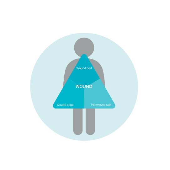
Wound Assessment form
PDF
Download the Triangle of Wound Assessment
2
pages
Before conducting a wound assessment, it's essential to recognise the signs of underlying arterial or venous disease. Understanding these indicators helps guide appropriate treatment decisions. For a deeper look at key risk factors and clinical signs to watch for, refer to the understanding leg ulcer section.
A full lower limb assessment, diagnosis and treatment plan should be completed within 14 days or in conjunction with your local policy.
Following a full lower limb assesment including an Ankle Brachial Pressure index, patients with suspected venous leg ulcers and adequate arterial supply, strong compression of 40mmHg therapy should be offered.
Personalised care is essential, including discussing causes, treatment options and supporting self‑management. Clear referral pathways should also be in place to allow direct access to relevant specialists without needing a GP referral.
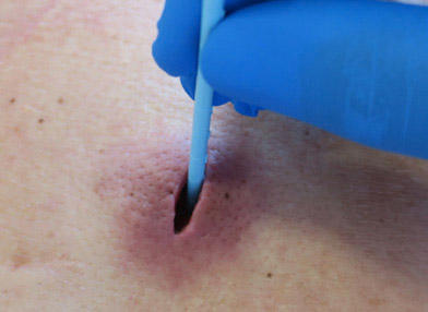
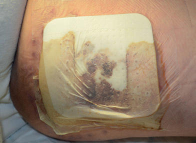
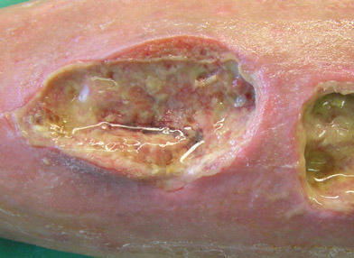
Assess the wound edge and periwound skin:
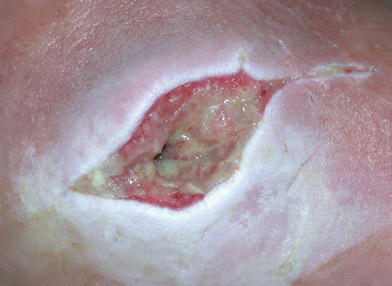
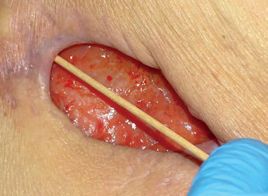
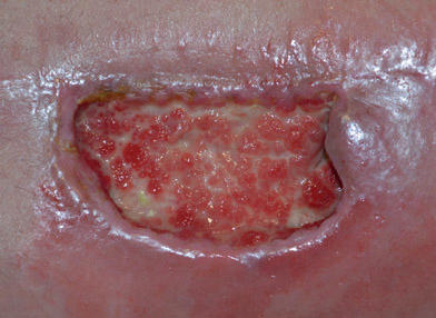
Wound Assessment can be complex with reasons for this being multifactorial. Using a simplified wound assessment framework such as the Triangle of Wound Assessment can help simplify the process.
Always look for signs of lymphoedema/lympho-venous disease as part of your clinical assessment
The Ankle Brachial Pressure Index (ABPI) is a quick, non‑invasive test that checks how well blood is flowing to the legs and feet.
It works by comparing two blood pressure readings:
Then the ankle pressure is divided by the arm pressure to give a number — the ABPI.
What the number tells you
Why it matters
Understanding ABPI helps clinicians decide:
ABPI values should always be interpreted in context of signs and symptoms.
Less than 0.5 suggests severe arterial disease
Between 0.8 and 1.3 suggests no evidence of significant arterial disease
Greater than 1.3 suggest arterial calcification
Greater than 0.5 to less than 0.8 suggests the presence of arterial disease or mixed arterial/venous disease
1. NICE (2021) How should I interpret ankle brachial pressure index (ABPI) results? Available at: Interpretation of ABPI | Diagnosis | Leg ulcer - venous | CKS | NICE
The aim of wound preparation is to accelerate healing or to facilitate the effectiveness of management and treatment plans. It is an approach that should be undertaken at each wound care intervention. When planning to prepare the wound you need to take into consideration:
Learn more about the importance of wound preparation, different debridement methods, and effectively using Alprep Pad for mechanical debridement.

Podiatry Operations Manager
Once your diagnosis of the leg has excluded arterial insufficiency, you are ready to determine the need for any additions to the standard treatment planning procedure.
Be prepared to manage exudate to protect the periwound skin, as venous leg ulcers are often highly exudating. (This will reduce over time with the correct level of compression therapy).
Frequently undertake a patient reported pain assessment, as leg ulcers are ranked as the most painful compared to other wounds.1
You should proceed according to the result of your ABPI:
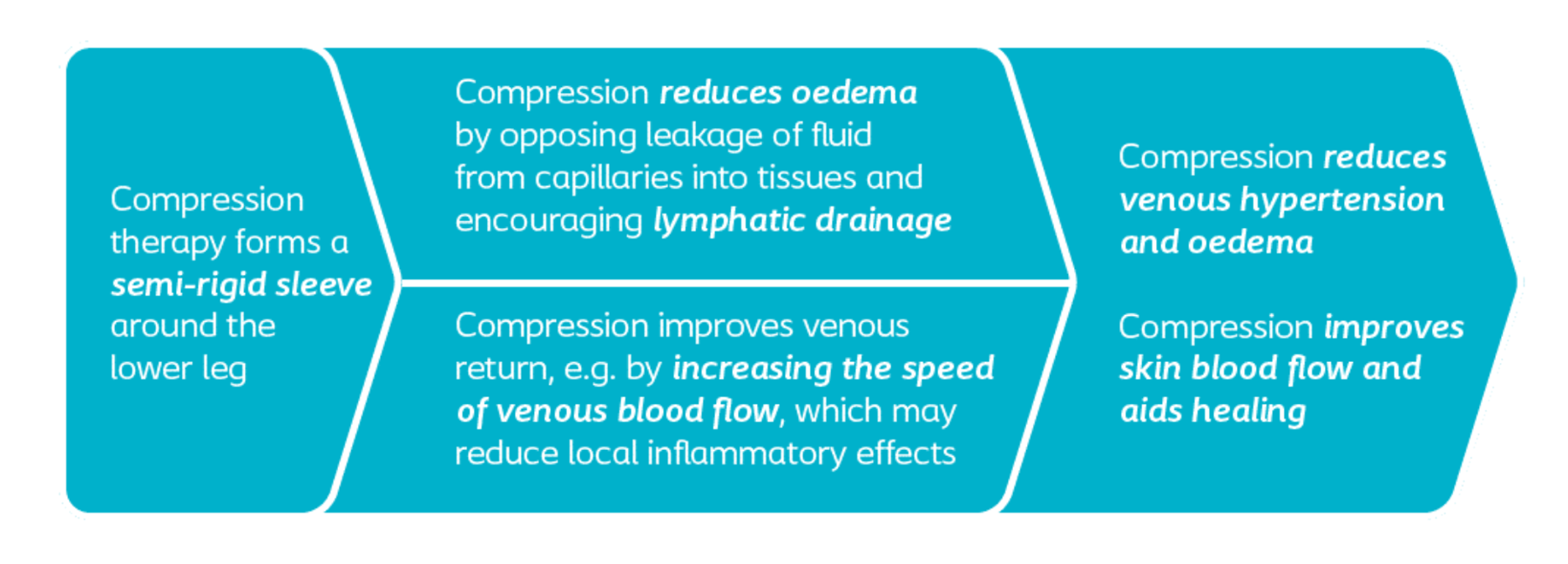
The concept of compression therapy
➔ Choose the correct type of compression by taking relevant factors into account: The severity of the disease, the anatomical characteristic of the leg/ankle, required frequency of applications, and the functionality of the patient: Patient preference, mobility, lifestyle and likely concordance.2
➔ Check for signs of infection at each dressing change, as infection and biofilm are common in venous leg ulcers. Use the IWII infection continuum and management guide.
Wound assessment and preparation leads to identification of objectives. We should aim to support an optimum healing environment for the wound.
Remember – Identifying and treatment of any underlying cause is integral to wound healing!
What is your treatment objective? Consider the following:
Management / prevention of...
The National Wound Care Strategy Programme recommends the application of a simple, low-adherent dressing with sufficient absorbency.
There are various dressing types available to us as clinicians, knowing what each does is essential.
Commonly used dressings can be broken down into categories:
)
Biatain Silicone with 3DFit Technology improves healing by conforming to the shape of the wound up to 2 cm deep*. This creates an optimal wound healing environment that reduces leakage along with the risk of maceration and infection.
*Tested in vitro, Conformability may vary across product design
)
Biatain® Silicone Non-Border consists of a soft and conformable polyurethane foam with a vapour permeable top film that is bacteria- and waterproof, a gentle and perforated silicone adhesive, and turquoise protective films.
)
Biatain® Non-Adhesive is a soft and conformable polyurethane foam dressing that effectively absorbs and retains wound exudate (1,2).
)
Biatain® Superabsorber is a soft, non-adhesive dressing that can manage high volumes of exudate, whilst protecting the wound edges and delicate peri-wound skin from maceration.
)
Biatain Silicone Ag is a conforming dressing with 3DFit Technology that fills the gap and reduces exudate pooling to promote optimal healing conditions.
)
Biatain® Ag Non-Adhesive can be used for a wide range of exuding wounds with delayed healing due to bacteria, or where there is a risk of infection
)
Biatain® Ibu Soft-Hold is a soft and flexible absorbent foam dressing containing ibuprofen and a gentle adherent layer covering less than 50% of the foam surface (1).
Hosiery kits for active ulcers: